Human anatomy is no doubt one of the first courses you must take in medical school. Whether you’re studying medicine, dentistry, optometry, nursing, or any other professional medical course, human anatomy is one of the basic medical subjects you’ll have before you can proceed through to the clinical classes.
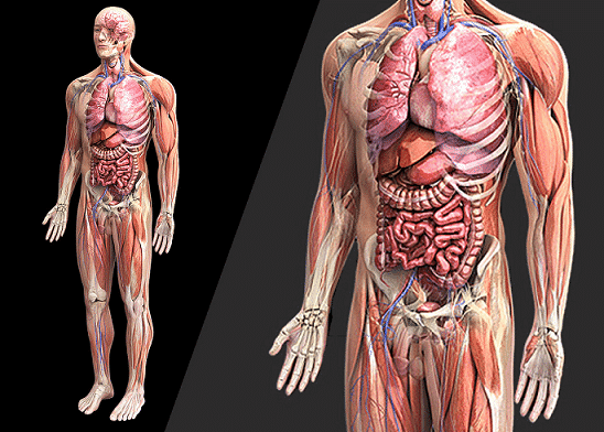
Now, in order to learn gross anatomy and understand it deeply, you’ll need more than just a good textbook on the subject. You’ll need to invest in a good atlas of human anatomy. While your textbook describes the various body structures and all you need to know about them, your atlas shows the real pictures of these structures, making your learning much easier.
DISCLOSURE: This post may contain affiliate links, meaning when you click the links and make a purchase, I receive a commission. As an Amazon Associate I earn from qualifying purchases.
When looking to buy an atlas of anatomy, there are so many options to choose from, and making the right choice can be a challenge. One of the biggest mistakes you can make at this stage is to choose a particular atlas because most of your colleagues are using the same or because your tutors have prescribed it. The truth is, each student is different, and the choice of atlas — as with any other book — should be based on personal learning styles and preferences.
Best Anatomy Atlases: Our Top 5 Picks
Here, we’ll be looking at some of the best and most authoritative human anatomy atlases you’ll find in the market. Our aim is to tell you, in brief, all you need to know about each option, so you can wind up with the right choice.
#1. Sobotta Atlas of Human Anatomy
![]()
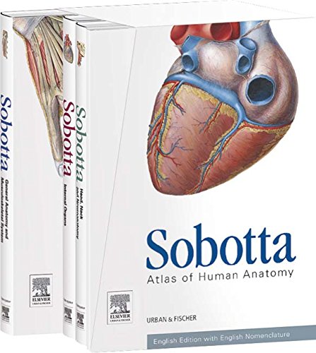
Though not the most widely known atlas on this list, we reckon that it’s the best you’ll find out there — for all the right reasons. This atlas is well organized, and the images are clear and detailed. Virtually all body structures are shown clearly from multiple views, and the labeling is clearly done. If you’re looking for images that are close to life-like, yet detailed enough to distinguish structures clearly, then you’d love this atlas.
Aside images of body structures, the atlas contains some text that briefly explain clinical correlations and help you to identify the most important features in each image. So, you can actually gather some good stuff while glancing through pages. And just so you know, the current edition comes in three volumes (Volume 1: General Anatomy & Musculoskeletal System, Volume 2: Internal Organs, Volume 3: Head Neck and Neuroanatomy). You know what that means? You’d have significantly less load to pack on your way to anatomy classes or study, since you only have to hold the volume you need.
- Authors: Friedrich Paulsen, Jens Waschke
- Publisher: Urban & Fischer
- Current edition: 15 edition (August 2013)
- Pages: 1,180
>>> Buy Now or Rent on Amazon.com
#2. Netter’s Atlas of Human Anatomy
![]()
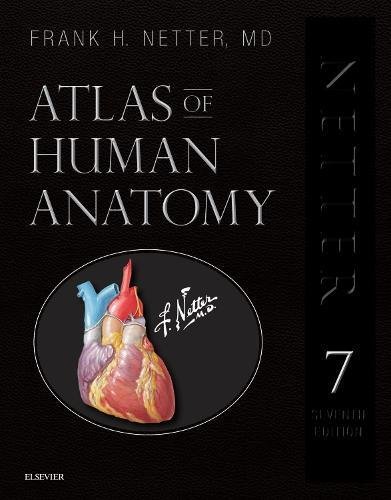
This is by far the most widely used human anatomy atlas in the country. Ask any medical or nursing student about it, and chances are they’ve seen or heard about it, at least. The atlas features bright and colorful images that show body structures in great detail. Although, the drawings look rather artificial, the coloring helps to distinguish structures clearly and makes them more memorable. If you’d love to practically visualize body structures in your head while on the go, then this atlas can help you achieve that.
Aside the artificial look of the drawings, there’s another downside: The pages contain only pictures; no texts to give additional information about structures. While some may argue that it’s just an atlas and you can always get all the information about each structure from your textbook, there are times when you won’t be with your textbook and you’d need some quick, short details about the structures you’re looking at.
- Author: Frank H. Netter MD
- Publisher: Elsevier
- Current edition: 7 edition (March 2018)
- Pages: 656
>>> Buy Now or Rent on Amazon.com
#3. Rohen’s Anatomy: A Photographic Atlas
![]()
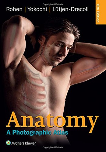
While this atlas isn’t as popular as the Netter’s, it’s more respected by those who have seen and compared both atlases. The biggest advantage of the Rohen’s atlas of anatomy is that it contains real life photographs. That is, rather than some artificial drawings that look too colorful to be true, you’d see the various body structures exactly as you’d find them in cadavers.
The atlas’s images are so crisp and life-like, you’d feel like you can touch and feel them on the pages. Note, however, that the atlas still uses colors where necessary to make distinctions that can easily be missed or confused.
If you want an atlas that will make your dissection sessions easier and more fun, you won’t go wrong with this. After all, the veins in the body aren’t blue and arteries aren’t red as depicted in some atlases. But it’s difficult to see some structures in striking detail because real photos are mostly used on the atlas’s pages. But then, your practical anatomy exams will be based on cadavers and cadaveric photos, not some crayon-garnished images.
- Authors: Johannes W. Rohen MD, Chihiro Yokochi MD, Elke Lütjen-Drecoll MD
- Publisher: LWW
- Current edition: Eighth, North American edition (February 2015)
- Pages: 560
>>> Buy Now or Rent on Amazon.com
#4. Gilroy’s Atlas of Anatomy (aka Thieme’s Atlas of Anatomy)
![]()
Authored by Anne Gilroy and few others and published by Thieme, this atlas of human anatomy is one of the options available in the market. It features pictures of super quality that are closer to real-life-like than being hand-drawn (they are computer-generated, though). Each body structure is shown in as much images as required for a good visual grasp of its details and relations. So, it strikes a balance between Netter’s obviously artificially colored images and the Rohen’s real-life images that are sometimes not-so-easy to appreciate.
As with our top pick (Sobotta’s atlas), the pages of this atlas include clinically oriented details rendered in colored boxes and tables. The text explains some of the most important details worth knowing about the structures shown on the page. And the current edition of the atlas includes a radiographs section, so you can visualize what each body structure looks like in X-rays and CT scans.
- Authors: Anne M Gilroy, Brian R MacPherson, Michael Schuenke, Erik Schulte, Udo Schumacher
- Publisher: Thieme
- Current edition: 3rd edition (April 2016)
- Pages: 760
>>> Buy Now or Rent on Amazon.com
#5. Clemente’s Anatomy: A Regional Atlas of the Human Body
![]()
If what you’re looking for is a trimmed-down version of the world-renowned Sobotta’s Atlas, then this atlas by Carmine Clemente is your best bet. It features over 1,000 clear, crisp, and realistically detailed color images of body structures, with accompanying diagnostic images and tables. The current edition also includes X-rays, CT scans, and sonograms, which are the clinically realistic non-invasive ways of viewing body structures.
If you think the Sobotta’s atlas isn’t well organized into volumes, then you’d find this a better alternative because its organization is just classical. And like some others on this list, the pages contain captions and text descriptions that provide additional information about the body structure shown. And more interestingly, the text in this atlas can be easily comprehended even by non-medical people.
- Author: Carmine D. Clemente PhD
- Publisher: LWW
- Current edition: Sixth, North American edition (February 2010)
- Pages: 752
There you have it — our list of the 5 best anatomy atlases. Whether you’re a preclinical student or even a resident, we believe that this post would have helped you decide on the right human anatomy atlas for you.
Related
- Best Books on Emergency Medicine (2022 Review)
- 20 Best Books on Diabetes Management (Type 1 & 2 – Pre-Diabetes Prevention)
- 20 Best Self Help Books For Depression And Anxiety
- 5 Best Medical Terminology Books for Beginners (2022 Review)
- 20 Best Books on Mindfulness for Beginners (Meditation & Positive Thinking)
- 20 Best Books on Meditation For Beginners (Transcendental, Mindfulness & Zen Meditation)
- 5 Best Anatomy Atlases Compared (2022 Review)
